Parts of the Heart: A Comprehensive Heart Glossary
Understanding the parts of the heart is crucial for comprehending how this vital organ functions to keep us alive. In this article, we’ll explore the anatomy of the heart and the role each part plays in maintaining circulatory health.
“Verified for accuracy, parts of the heart listed here have been sourced from reputable references. Source: Your Info Master.”
- Parts of the Heart
- Cardiac Glossary
- Aorta
- Aortic Valve
- Arrhythmia
- Artery
- Atresia
- Atria (singular: atrium)
- AV (Atrioventricular) Node
- Balloon Valvuloplasty
- Bicuspid
- Biological Valve
- Blood Vessels
- Bradycardia or Bradyarrhythmia
- Bundle Branches
- Cardio
- Cardiologist
- Cardiomyopathy
- Catheter
- Coarctation
- Contrast Dye
- Coronary
- Coronary Arteries
- Cyanosis
- Ductus Arteriosus
- Echocardiography (Echo)
- Edema
- Electrocardiography (ECG or EKG)
- Foramen Ovale
- Homograft
- Hypertrophy
- Hypoplastic
- Insufficiency (Regurgitation)
- IV (Intravenous) Line
- Mechanical Valve
- Mitral Valve
- Murmur
- Palpitation
- Pulmonary Artery
- Pulmonary Veins
- Pulmonary Valve
- Risk Factors
- SA (Sinoatrial) Node
- Shunt
- Stenosis
- Stent
- Tachycardia or Tachyarrhythmia
- Tricuspid Valve
- Valves
- Vein
- Ventricles (singular: Ventricle)
- What are the valves of the heart?
- Conclusion
- Related Articles
Read also: Body Parts Names
Parts of the Heart
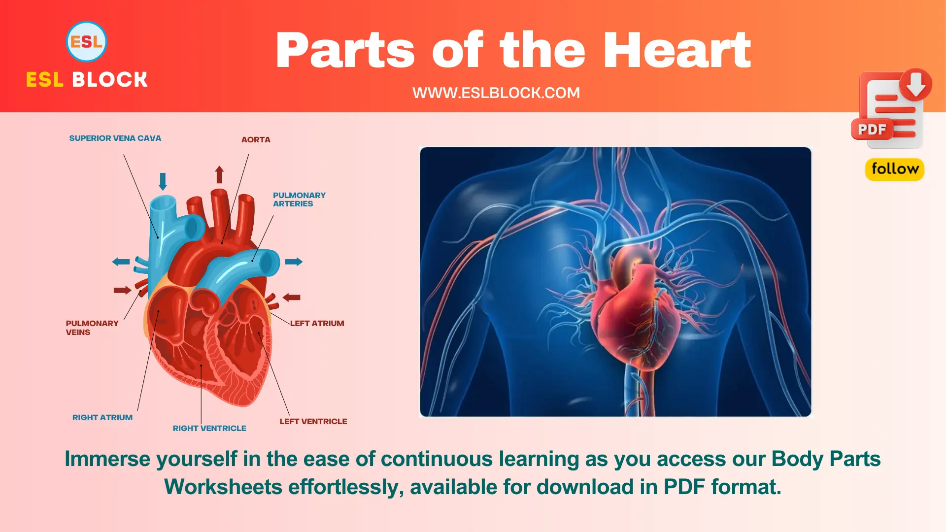
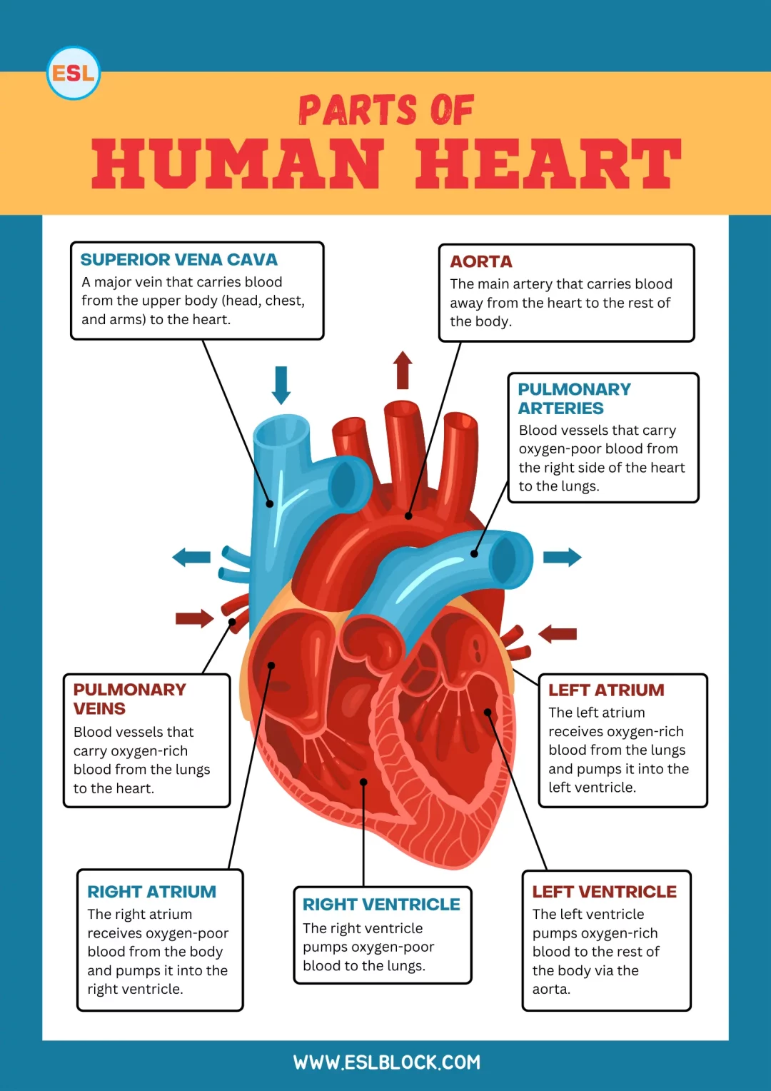
The human heart is a complex organ with several key parts, each with specific functions. Here are the main parts of the human heart:
- Atria: The two upper chambers of the heart.
- Right Atrium: Receives deoxygenated blood from the body.
- Left Atrium: Receives oxygenated blood from the lungs.
- Ventricles: The two lower chambers of the heart.
- Right Ventricle: Pumps deoxygenated blood to the lungs.
- Left Ventricle: Pumps oxygenated blood to the rest of the body.
- Valves: Ensure one-way blood flow through the heart.
- Tricuspid Valve: Between the right atrium and right ventricle.
- Pulmonary Valve: Between the right ventricle and pulmonary artery.
- Mitral (bicuspid) Valve: Between the left atrium and left ventricle.
- Aortic Valve: Between the left ventricle and aorta.
- Septum: The wall separating the heart’s left and right sides.
- Interatrial Septum: Separates the atria.
- Interventricular Septum: Separates the ventricles.
- Blood Vessels: Major vessels associated with the heart.
- Aorta: The main artery carrying oxygenated blood from the left ventricle to the body.
- Pulmonary Arteries: Carry deoxygenated blood from the right ventricle to the lungs.
- Pulmonary Veins: Carry oxygenated blood from the lungs to the left atrium.
- Superior and Inferior Vena Cava: Bring deoxygenated blood from the body to the right atrium.
- Coronary Arteries: Supply blood to the heart muscle itself.
- Chordae Tendineae: Tendinous cords that connect the valves to the heart muscles, preventing valve prolapse.
- Papillary Muscles: Muscles located in the ventricles that anchor the chordae tendineae.
Also Check: 25 Chili Pepper Types
Cardiac Glossary
Aorta
The body’s largest artery. It carries oxygen-rich blood from the heart to the rest of the body.
Aortic Valve
A valve inside the heart that allows blood to flow forward from the left ventricle to the aorta.
Arrhythmia
An abnormal heart rhythm or rate.
Artery
A blood vessel that carries blood away from the heart.
Atresia
A condition in which a part of the heart, such as a valve, is absent at birth because it didn’t develop correctly.
Atria (singular: atrium)
The heart’s two upper chambers. They receive blood from the lungs (left atrium) and the body (right atrium).
AV (Atrioventricular) Node
The cluster of electrical cells in the heart receives signals from the atria and guides them to the ventricles.
Balloon Valvuloplasty
A procedure that uses a balloon-tipped catheter to open a narrowed heart valve or vessel.
Bicuspid
A heart valve with two leaflets.
Biological Valve
A heart valve is created from human or animal tissue.
Blood Vessels
Tubes that carry blood throughout the body. Arteries and veins are blood vessels.
Bradycardia or Bradyarrhythmia
A type of arrhythmia occurs when the heart beats too slowly.
Bundle Branches
Pathways of cells in the heart that carry electrical signals from the AV node into the ventricles.
Cardio
Relating to the heart.
Cardiologist
A doctor with special training to diagnose and treat heart problems.
Cardiomyopathy
Structural and/or functional diseases of the heart’s ventricles are not caused by coronary artery disease.
Catheter
A long, thin, flexible tube is used during cardiac catheterization procedures to obtain information about the heart or treat a heart problem.
Coarctation
Narrowing of a blood vessel.
Contrast Dye
A special fluid that enhances x-rays of blood vessels, allowing blood flow to be tracked. Used for certain heart tests.
Coronary
Relating to vessels that provide blood to the heart itself.
Coronary Arteries
Blood vessels that wrap around the heart and supply the heart muscle with oxygen-rich blood.
Cyanosis
A condition in which the skin, lips, and nails appear blue due to low oxygen levels in the blood.
Ductus Arteriosus
A blood vessel in the fetus that connects the pulmonary artery and the aorta. If it fails to close after birth, it’s called a patent ductus arteriosus (PDA).
Echocardiography (Echo)
A test that uses sound waves to create a picture of the heart. Also called a heart ultrasound.
Edema
A buildup of excess fluid in the body, often shows up as swollen feet or ankles.
Electrocardiography (ECG or EKG)
A test that records the way electrical signals move through the heart.
Foramen Ovale
An opening between the two upper chambers of a fetus’s heart. If it fails to close after birth, it’s called a patent foramen ovale (PFO).
Homograft
A blood vessel with or without a heart valve from a human donor.
Hypertrophy
When the heart muscle thickens.
Hypoplastic
Abnormally small or undeveloped.
Insufficiency (Regurgitation)
When a heart valve doesn’t close tightly, allowing leakage or backward flow of blood.
IV (Intravenous) Line
A thin tube that delivers fluid to a vein.
Mechanical Valve
A heart valve is created from man-made materials, including ceramic or metal.
Mitral Valve
The valve inside the heart allows blood to flow forward from the left atrium to the left ventricle.
Murmur
The heart makes an extra noise when blood doesn’t flow smoothly through it.
Palpitation
An irregular or skipped heartbeat.
Pulmonary Artery
The large artery that carries blood from the heart to the lungs to get oxygen.
Pulmonary Veins
Veins that carry oxygen-rich blood from the lungs to the heart.
Pulmonary Valve
The valve inside the heart allows blood to flow forward from the right ventricle to the pulmonary artery.
Risk Factors
Behaviors and/or conditions that place a person at higher risk of having a problem or disease.
SA (Sinoatrial) Node
A cluster of electrical cells in the right atrium starts each heartbeat.
Shunt
When blood flows from one location of the heart to another in the path of least resistance, it can also refer to a tube or device placed in the heart to allow blood to flow in a certain direction.
Stenosis
Narrowing at or near a heart valve or blood vessel obstructs blood flow.
Stent
A device is placed in a blood vessel or heart valve to help keep it open.
Tachycardia or Tachyarrhythmia
A type of arrhythmia occurs when the heart beats too fast.
Tricuspid Valve
The valve inside the heart allows blood to flow forward from the right atrium to the right ventricle.
Valves
“Doorways” that open and close to allow blood to flow forward through the heart.
Vein
A blood vessel that carries blood toward the heart.
Ventricles (singular: Ventricle)
The heart’s two lower chambers pump blood to the lungs (right ventricle) and the body (left ventricle).
What are the valves of the heart?
The heart has four valves to ensure that blood only flows in one direction:
Aortic Valve
This valve is located between the left ventricle and the aorta.
Mitral Valve
This valve is located between the left atrium and the left ventricle.
Pulmonary Valve
This valve is located between the right ventricle and the pulmonary artery.
Tricuspid Valve
This valve is located between the right atrium and the right ventricle.
Most people are familiar with the sound of the heart. In fact, the heart makes many types of sounds, and doctors can distinguish these to monitor the heart’s health.
The opening and closing of the valves are key contributors to the sound of the heartbeat. If there is leaking or a blockage of the heart valves, it can create sounds called “murmurs.”
Conclusion
By familiarizing yourself with the different parts of the heart, you gain a deeper appreciation for its intricate design and essential functions. Stay informed about heart health to ensure your cardiovascular system remains strong and efficient.
If you have enjoyed “Parts of the Heart,” I would be very thankful if you could help spread the word by emailing it to your friends or sharing it on Twitter, Instagram, Pinterest, or Facebook. Thank you!
ESLBLOCK will teach you new things. If you have questions about the major parts of the heart, you can ask us in the comments section of our blog. Come back often! We will try to answer you quickly. Thank you!
Also Read: Parts of the Face with Pictures
Related Articles
Here are some more lists for you!
- Bone Names of the Human Skeleton
- Names of Teeth in English with Pictures
- Parts of the Mouth Vocabulary
- Parts of the Hand Vocabulary

